a

Vision and Vision Center Clear Vision 12 years of experience and all doctors who conduct operations and supervise the cases professors in the universities of Egypt The use of the latest German technology for eye correction and examination devices in the world is the first Egyptian center collaborates with the Center Vysium in Spain and the only center in Egypt that owns the Corvis ST is the most accurate device in the world to measure the strength of the cornea of the eye to determine the appropriate operation by 100%

+2 01224247777

info@clearvisionlasik.net
54 El Batal Ahmed Abdel Aziz St., Mohandeseen, Giza

Recent Posts

Follow Us On Instagram

Saturday - Thursday 9.00 - 6.00

Friday - Holiday

+2 01224247777

info@clearvisionlasik.net

Center Location

54 El Batal Ahmed Abdel Aziz St, Giza

Follow Us

Book An Appointment

Clear Vision Laser Center  >  Separation Retinal

Separation Retinal

What does retinal detachment mean?

Retinal detachment occurs when the inner lining of the eye (retina) separates from the eye wall (placenta), which contains blood vessels that supply the eye with oxygen and nutrients. The fluid accumulates between the two layers, which affects retinal cells and weakens vision.

There are 3 different types of retinal detachment as follows

Rhegmatogenous retinal detachment: It occurs as a result of holes or gap in the retina which allows the passage of fluid under the retina and separating it from the placenta as a result of this rupture.
Exudative Retinal detachment: An inflammatory disorder that causes the accumulation of fluid behind the retina.
Trancinal Retinal detachment occurs when fibrous tissue is formed to tighten the retina.


Retinal detachment symptoms


Sudden appearance of black spots, spots or lines floating within the field of vision.
Seeing a large number of floating objects, some may notice one large black shape floating within the field of vision such as a fly.
Sudden short flashes of light not exceeding one second appear in front of the infected eye.
A dark shadow vision appears as a curtain from the outside side of the field of vision in the affected eye.
Sometimes confusing vision.


Causes of retinal detachment


Age in which retinal detachment is more common in people over the age of 50.
Retinal detachment occurs in one eye.
Family history of retinal detachment.
Severe myopia.
Exposure to previous eye surgery, such as removal of cataracts.
Exposure to disease or other eye inflammation before.
Diabetics are more symptomatic of retinal detachment.


Diagnosis


Your doctor may use the following tests to diagnose retinal detachment:

Check out a natural look to measure your vision and ability to distinguish colors.
Retinal examination by using a tool with bright light and special lenses to examine the back of the eye, including the retina.
Ultrasonic imaging is used if bleeding occurs in the eye, making it difficult to see the retina.
Check the eye pressure and blood flow throughout your eye and specifically the retina.
Your doctor may also test the ability of the retina to send nerve signals to your brain.


Retinal detachment treatment

Retinal detachment is necessary because one of the important consequences of retinal detachment is the irreversible loss of vision due to retinal atrophy or chronic osteoarthritis, which causes the entire eye to become obese.

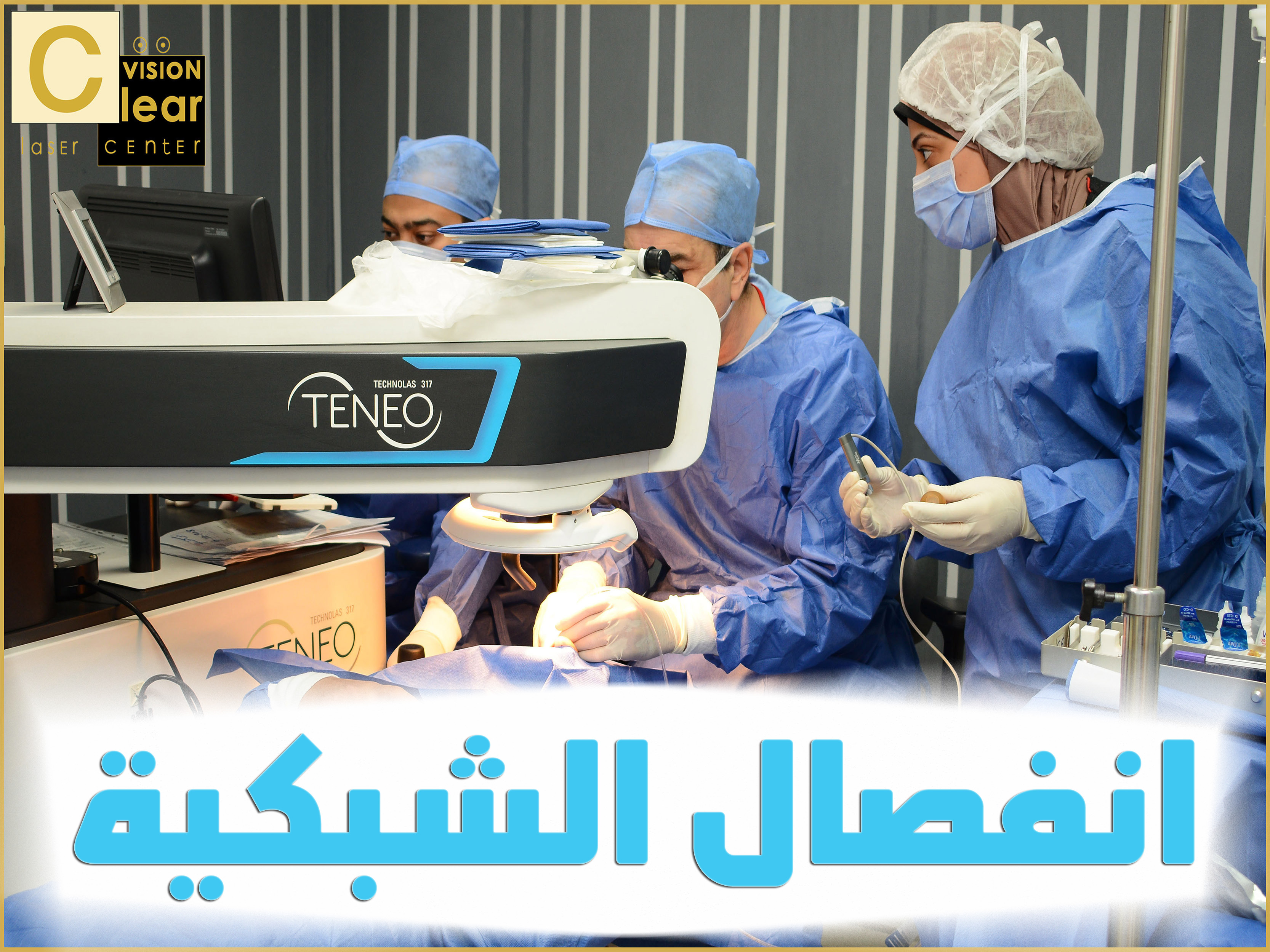
Surgical treatment is always a choice to repair ruptures and holes that cause retinal detachment by the laser or cold retinal probe to retract the retina by pushing the back wall of the eye from the outside toward the retina.

This is done by suspending ribbons or parts of silicon or removing the glass and inserting gas or silicon oil into the glass cavity. The operation is performed under the influence of the local anesthetic and does not need to be kept in the hospital.

admin
No Comments

Leave a Reply